Temporalis Muscle Cavernous Hemangioma in a Pediatric Patient
DOI:
https://doi.org/10.46900/apn.v7i2.286Keywords:
temporalis, hemangioma, cavernous hemangioma, intramuscular hemangioma, pediatricAbstract
Background: Temporalis muscle hemangiomas are an uncommon entity, even among the rare incidences of intramuscular hemangiomas. They are frequently identified as round, painless, soft lesions beneath the skin that have been growing slowly over time. A preliminary diagnosis can be made based on magnetic resonance imaging, but it is confirmed by pathology of the surgically excised lesion. The safest practice is to resect all of the hemangioma plus a safe margin of normal muscle so as to prevent recurrence as much as possible. Watchful waiting is also a reasonable alternative when surgery is not indicated.
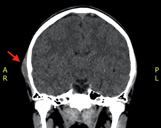
Case presentation: Because of the apparent paucity of this lesion, especially in the pediatric population, we report this case of a temporalis muscle cavernous hemangioma in a male under 18 years of age who presented for a growing, painful temporalis lesion 4 years after being managed conservatively.
Conclusion: Temporalis hemangiomas are among the rarest intramuscular hemangiomas (IMHs) reported in the pediatrics population. They are often misdiagnosed, with MRIs being the preferred imaging for preoperative diagnosis, and a CT can determine bony involvement. The superior method of treatment when not simply observing its progress is wide surgical excision with normal muscle borders preceded by embolization for better bleeding control. IMHs do not have very high recurrence rates, but re-operation is feasible and none was shown to metastasize.
Downloads
Downloads
Published
How to Cite
Issue
Section
Categories
License
Copyright (c) 2025 Asmaa Kebbe, Mohamad Nabih El Houshiemy, Khaled Sidani, Sarah Kawtharani, Marwan Najjar

This work is licensed under a Creative Commons Attribution 4.0 International License.

When publishing in Archives of Pediatric Neurosurgery journal, authors retain the copyright of their article and agree to license their work using a Creative Commons Attribution 4.0 International Public License (CC BY 4.0), thereby accepting the terms and conditions of this license (https://creativecommons.org/licenses/by/4.0/legalcode).
The CC BY 4.0 license terms applies to both readers and the publisher and allows them to: share (copy and redistribute in any medium or format) and adapt (remix, transform, and build upon) the article for any purpose, even commercially, provided that appropriate credit is given to the authors and the journal in which the article was published.
Authors grant Archives of Pediatric Neurosurgery the right to first publish the article and identify itself as the original publisher. Under the terms of the CC BY 4.0 license, authors allow the journal to distribute the article in third party databases, as long as its original authors and citation details are identified.