Imaging features on Chiari 0 malformation, is there any difference with Chiari I? A clinical image.
DOI:
https://doi.org/10.46900/apn.v4i2(May-August).98Keywords:
Chiari 0, Chiari I, Magnetic resonance, Posterior fossa, syringohydromyelia, Chiari malformationAbstract
Distinction between Chiari 0 and Chiari I malformation on MRI is difficult; both diseases share a common pathophysiology, and some theories explain that them constitute a continuum rather than distinct malformations (1).
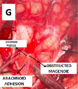
CM0 malformation was first described in 1998 (2), as a classic syringohydromyelia with no or minimal tonsillar herniation (<3mm), crowding of the foramen magnum and obstruction of the Magendie by adhesions (3)
This case of a 3-year–old patient with occipital headaches, neck pain, and sleep apneas with a cervicothoracic syringohydromyelia on MRI had a complete improvement of his symptoms after a suboccipital craniectomy, removal of 2/3 of the posterior arch of C1 and release of the choroid plexus and arachnoid adhesions on the foramen of Magendie. Other anatomical variants (also seem on MRI) include tips of the obices located below normal position, increasing of the anteroposterior midsagittal distance of the spinomedullary junction and an increased angle between the clivus an the fourth ventricle (4), making a diagnosis of Chiari 0 malformation on this clinical image.
Obstructed CSF flow without tonsillar herniation could be attributable to the compactness of the posterior fossa with arachnoidal adhesions at the fourth ventricular outlet (5.)
Downloads
References
1.Azahraa Haddad F, Qaisi I, Joudeh N, Dajani H, Jumah F, Elmashala A, Adeeb N, Chern JJ, Tubbs RS. The newer classifications of the chiari malformations with clarifications: an anatomical review. Clinical Anatomy. 2018 Apr;31(3):314-22.
2.Iskandar BJ, Hedlund GL, Grabb PA, Oakes WJ. The resolution of syringohydromyelia without hindbrain herniation after posterior fossa decompression. Journal of neurosurgery. 1998 Aug 1;89(2):212-6.
3. Cesmebasi A, Loukas M, Hogan E, Kralovic S, Tubbs RS, Cohen?gadol AA. The C hiari malformations: A review with emphasis on anatomical traits. Clinical anatomy. 2015 Mar;28(2):184-94.
4. Tubbs RS, Elton S, Grabb P, Dockery SE, Bartolucci AA, Oakes WJ. Analysis of the posterior fossa in children with the Chiari 0 malformation. Neurosurgery. 2001 May 1;48(5):1050-5.
5. Kyoshima K, Kuroyanagi T, Oya F, Kamijo Y, El-Noamany H, Kobayashi S. Syringomyelia without hindbrain herniation: tight cisterna magna: report of four cases and a review of the literature. Journal of Neurosurgery: Spine. 2002 Mar 1;96(2):239-49.
Additional Files
Published
How to Cite
Issue
Section
License
Copyright (c) 2021 Felipe Gutierrez Pineda

This work is licensed under a Creative Commons Attribution 4.0 International License.

When publishing in Archives of Pediatric Neurosurgery journal, authors retain the copyright of their article and agree to license their work using a Creative Commons Attribution 4.0 International Public License (CC BY 4.0), thereby accepting the terms and conditions of this license (https://creativecommons.org/licenses/by/4.0/legalcode).
The CC BY 4.0 license terms applies to both readers and the publisher and allows them to: share (copy and redistribute in any medium or format) and adapt (remix, transform, and build upon) the article for any purpose, even commercially, provided that appropriate credit is given to the authors and the journal in which the article was published.
Authors grant Archives of Pediatric Neurosurgery the right to first publish the article and identify itself as the original publisher. Under the terms of the CC BY 4.0 license, authors allow the journal to distribute the article in third party databases, as long as its original authors and citation details are identified.