Endoscopic anatomy of the ventricles
DOI:
https://doi.org/10.46900/apn.v5Suppl1.132Keywords:
Neuroendoscopy, anatomyAbstract
The cerebral ventricles are structures known since antiquity. At the beginning of its description there was a belief that human thoughts and intellect were generated inside the ventricular cavity, and this idea persisted even after the Renaissance. In this period after the Dark Ages, the anatomical description of the ventricles evolved a lot, but it took a long time to conclude the true liquid nature of the ventricular content. The ventricles are divided into lateral, third and fourth ventricles, and each of these regions presents communications between themselves and very precise limits. For endoscopic procedures, a thorough knowledge of the anatomy of the lateral and third ventricles is extremely important. The cerebrospinal fluid circulates inside the ventricles according to a complex mechanism, and there are still controversies nowadays, especially regarding its resorption.
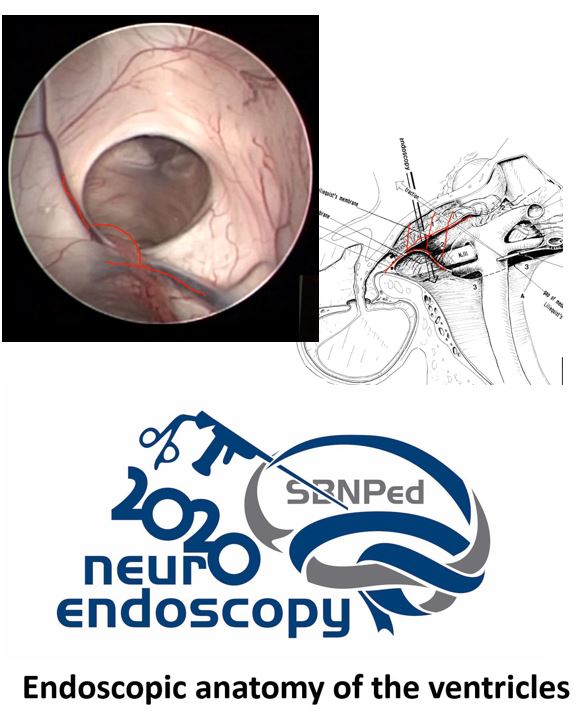
Each lateral ventricle is a “C”-shaped cavity that surrounds the thalamus and is deeply located in the brain, following the contour of the choroid fissure. These structures represent, from an embryological point of view, the light of the telencephalic vesicles. Each lateral ventricle has five parts: frontal horn, body, atrium, occipital horn, and temporal horn. The frontal horn is the part of the lateral ventricle located anteriorly to the foramen of Monro. The lateral ventricle body extends from the posterior margin of the foramen of Monro to the point where the fornix and the corpus callosum converge, thus disappearing the septum pellucidum. The atrium of the lateral ventricle and its occipital extension form a triangle with an anterior base in the pulvinar and a posterior apex in the occipital lobe. The occipital horn is triangular, with a posterior vertex and a base in the atrium. The temporal horn is the inferior extension of the ventricle, being the continuation of the atrium, directing itself anteriorly and laterally. Each of these divisions has a medial, lateral, ceiling, and floor wall. In addition, the frontal, temporal, and atrial horns have anterior walls. These walls are formed by the thalamus, septum pellucidum, white matter, corpus callosum, caudate nucleus, and fornix. The third ventricle is a funnel-shaped, unilocular, narrow midline cavity. It communicates at the anterosuperior margin with each lateral ventricle through the foramen of Monro and, subsequently, with the fourth ventricle through the cerebral aqueduct. In adult individuals, the lateral distance of the third ventricle is 5.5 mm on average. In a study using MRI images, the hydrocephalic configuration of the third ventricle disappeared after the endoscopic third ventriculostomy (ETV), with a decrease in diameter, elevation, and horizontal direction of the floor and reduction of the infundibular angle . The floor of the third ventricle extends from the optic chiasm anteriorly to the opening of the cerebral aqueduct posteriorly. It descends ventral and is formed by at least 12 cellular clusters or nuclei within the hypothalamic region. Anatomically, three portions can be described on the floor of the third ventricle: (1) premammillary portion, which extends from the infundibulum to the premammillary sulcus, constituting a very thin layer of gray substance of the hypothalamus; (2) interpeduncular portion, which extends from the postmammillary recess to the posterior margin of the interpeduncular space, being formed of gray substance and firmer than the first; and (3) peduncular portion, which corresponds to the portion of the cerebral peduncles, being the most solid portion, being formed by the medial aspect of the peduncles covered by the peduncular ependyma.
The knowledge of the endoscopic anatomy of the ventricles is of the key importance for endoscopic neuroendoscopy
Downloads
References
Dezena RA. Atlas of Endoscopic Neurosurgery of the Third Ventricle. Basic Principles for Ventricular Approaches and Essential Intraoperative Anatomy. 1ed. Cham, Suiça: Springer International Publishing, 2017. v. 1. 271p. doi: 10.1007/978-3-319-50068-3
Dezena RA. Endoscopic Third Ventriculostomy. Classic Concepts and a State-of-the-Art Guide. 1ed. Cham, Suiça: Springer International Publishing, 2020. v. 1. 111p. doi: 10.1007/978-3-030-28657-6
Downloads
Additional Files
Published
How to Cite
Issue
Section
License
Copyright (c) 2022 Roberto Alexandre Dezena

This work is licensed under a Creative Commons Attribution 4.0 International License.

When publishing in Archives of Pediatric Neurosurgery journal, authors retain the copyright of their article and agree to license their work using a Creative Commons Attribution 4.0 International Public License (CC BY 4.0), thereby accepting the terms and conditions of this license (https://creativecommons.org/licenses/by/4.0/legalcode).
The CC BY 4.0 license terms applies to both readers and the publisher and allows them to: share (copy and redistribute in any medium or format) and adapt (remix, transform, and build upon) the article for any purpose, even commercially, provided that appropriate credit is given to the authors and the journal in which the article was published.
Authors grant Archives of Pediatric Neurosurgery the right to first publish the article and identify itself as the original publisher. Under the terms of the CC BY 4.0 license, authors allow the journal to distribute the article in third party databases, as long as its original authors and citation details are identified.